From Wikipedia, the free encyclopedia
| Pineal gland | |
|---|---|
 | |
| Diagram of pituitary and pineal glands in the human brain | |
| Latin | glandula pinealis |
| Gray's | subject #276 1277 |
| Artery | posterior cerebral artery |
| Precursor | Neural Ectoderm, Roof of Diencephalon |
| MeSH | Pineal+gland |
Contents[hide] |
[edit] Location
The pineal gland is reddish-gray and about the size of a grain of rice (5–8 mm) in humans, located just rostro-dorsal to the superior colliculus and behind and beneath the stria medullaris, between the laterally positioned thalamic bodies. It is part of the epithalamus.The pineal gland is a midline structure shaped like a pine cone,[3] and is often seen in plain skull X-rays, as it is often calcified;[4] calcification has been shown in one study to correlate with the accumulation of fluoride.[5]
[edit] Structure and composition


The pineal gland consists mainly of pinealocytes, but four other cell types have been identified. As it is quite cellular (in relation to the cortex and white matter) it may be mistaken for a neoplasm.[6]
| Cell type | Description |
|---|---|
| Pinealocytes | The pinealocytes consist of a cell body with 4–6 processes emerging. They produce and secrete melatonin. The pinealocytes can be stained by special silver impregnation methods. Their cytoplasm is lightly basophilic. With special stains, pinealocytes exhibit lengthy, branched cytoplasmic processes which extend to the connective septa and its blood vessels. |
| Interstitial cells | Interstitial cells are located between the pinealocytes. They have elongated nuclei and a cytoplasm which is stained darker than that of the pinealocytes. |
| Perivascular phagocyte | Many capillaries are present in the gland, and perivascular phagocytes are located close to these blood vessels. The perivascular phagocytes are antigen presenting cells. |
| pineal neurons | In higher vertebrates neurons are located in the pineal gland. However, these are not present in rodents. |
| peptidergic neuron-like cells | In some species, neuronal-like peptidergic cells are present. These cells might have a paracrine regulatory function. |
[edit] Miscellaneous anatomy
Pinealocytes in many non-mammalian vertebrates have a strong resemblance to the photoreceptor cells of the eye. Some evolutionary biologists believe that the vertebrate pineal cells share a common evolutionary ancestor with retinal cells.[10]In some vertebrates, exposure to light can set off a chain reaction of enzymatic events within the pineal gland that regulate circadian rhythms.[11] Some early vertebrate fossil skulls have a pineal foramen (opening). This correlates with the physiology of the modern "living fossils," the lamprey and the tuatara, and some other vertebrates that have a parietal organ or "third eye," which, in some of them, is photosensitive. The third eye represents evolution's earlier approach to photoreception.[12] The structures of the third eye in the tuatara are analogous to the cornea, lens and retina, though the latter resembles that of an octopus rather than a vertebrate retina. The asymmetrical whole consists of the "eye" to the left and the pineal sac to the right. "In animals that have lost the parietal eye, including mammals, the pineal sac is retained and condensed into the form of the pineal gland."[12]
Unlike much of the rest of the mammalian brain, the pineal gland is not isolated from the body by the blood–brain barrier system;[13] it has profuse blood flow, second only to the kidney.
Fossils seldom preserve soft anatomy. The brain of the Russian Melovatka bird, about 90 million years old, is an exception, and it shows a larger-than-expected parietal eye and pineal gland.[14]
In humans and other mammals, the light signals necessary to set circadian rhythms are sent from the eye through the retinohypothalamic system to the suprachiasmatic nuclei (SCN) and the pineal.
[edit] Function
The pineal gland was originally believed to be a "vestigial remnant" of a larger organ. In 1917 it was known that extract of cow pineals lightened frog skin. Dermatology professor Aaron B. Lerner and colleagues at Yale University, hoping that a substance from the pineal might be useful in treating skin diseases, isolated and named the hormone melatonin in 1958.[15] The substance did not prove to be helpful as intended, but its discovery helped solve several mysteries such as why removing the rat's pineal accelerated ovary growth, why keeping rats in constant light decreased the weight of their pineals, and why pinealectomy and constant light affect ovary growth to an equal extent; this knowledge gave a boost to the then new field of chronobiology.[16]Melatonin is N-acetyl-5-methoxy-tryptamine, a derivative of the amino acid tryptophan, which also has other functions in the central nervous system. The production of melatonin by the pineal gland is stimulated by darkness and inhibited by light.[17] Photosensitive cells in the retina detect light and directly signal the SCN, entraining its rhythm to the 24-hour cycle in nature. Fibers project from the SCN to the paraventricular nuclei (PVN), which relay the circadian signals to the spinal cord and out via the sympathetic system to superior cervical ganglia (SCG), and from there into the pineal gland.
The compound pinoline is also produced in the pineal gland; it is one of the beta-carbolines.[citation needed]
The human pineal gland grows in size until about 1–2 years of age, remaining stable thereafter,[18][19] although its weight increases gradually from puberty onwards.[20][21] The abundant melatonin levels in children are believed to inhibit sexual development, and pineal tumors have been linked with precocious puberty. When puberty arrives, melatonin production is reduced.
Calcification of the pineal gland is typical in adults, and has been observed in children as young as 2. Calcification rates vary widely by country and tend to increase by age, with calcification occurring in an estimated 40% of Americans by their 17th year.[4]
Apparently the internal secretions of the pineal gland inhibit the development of the reproductive glands, because in cases where it is severely damaged in children, the result is accelerated development of the sexual organs and the skeleton.[22] In animals, the pineal gland appears to play a major role in sexual development, hibernation, metabolism, and seasonal breeding.[23]
Pineal cytostructure seems to have evolutionary similarities to the retinal cells of chordates.[10] Modern birds and reptiles have been found to express the phototransducing pigment melanopsin in the pineal gland. Avian pineal glands are believed to act like the SCN in mammals.[24]
Studies on rodents suggest that the pineal gland may influence the actions of recreational drugs, such as cocaine,[25] and antidepressants, such as fluoxetine (Prozac),[26] and its hormone melatonin can protect against neurodegeneration.[27]
[edit] Conjecture
Dr. Rick Strassman, while conducting research on the psychedelic dimethyltryptamine (DMT) in the 1990s at the University of New Mexico, advanced the controversial hypothesis that a massive release of DMT from the pineal gland prior to death or near death was the cause of the near death experience (NDE) phenomenon. Several of his test subjects reported NDE-like audio or visual hallucinations. His explanation for this was the possible lack of panic involved in the clinical setting and possible dosage differences between those administered and those encountered in actual NDE cases. Several subjects also reported contact with 'other beings', alien like, insectoid or reptilian in nature, in highly advanced technological environments[28] where the subjects were 'carried,' 'probed,' 'tested,' 'manipulated,' 'dismembered,' 'taught,' 'loved,' and even 'raped' by these 'beings' (one could note the strong similarities of these bodily tests/invasions in other psychedelic experiences throughout time, outlined in Graham Hancock's "Supernatural"[29]). Basing his reasoning on his belief that all the enzymatic material needed to produce DMT is found in the pineal gland (see evidence in mammals), and moreover in substantially greater concentrations than in any other part of the body, Strassman ([28] p. 69) has speculated that DMT is made in the pineal gland.[edit] Pathology
All tumors involving the pineal gland are rare; most (50% to 70%) arise from sequestered embryonic germ cells. They most commonly take the form of so-called germinomas, resembling testicular seminoma or ovarian dysegerminoma. Other lines of germ cell differentiation include embryonal carcinomas; choriocarcinomas; mixtures of germinom, embryonal carcinoma, and choriocarcinoma; and, uncommonly, typical teratomas (usually benign). Whether to characterize these germ cell neoplasms as pinealomas is still a subject of debate, but most pinealophiles favor restricting the terms pinealoma to neoplasms arising from the pineocytes.A pineal tumor can compress the superior colliculi and pretectal area of the dorsal midbrain, producing Parinaud's syndrome. Pineal tumors also can cause compression of the cerebral aqueduct, resulting in a noncommunicating hydrocephalus.
[edit] Pinelomas
These neoplasms are divided into two categories, pineoblastomas and pineocytomas,[citation needed] based on their level of differentiation, which in turn, correlates with their neoplastic aggressiveness. The clinical course of patients with pineocytomas is prolonged, averaging 7 years.[citation needed] The manifestations are the consequence of their pressure effects and consist of visual disturbances, headache, mental deterioration, and sometimes dementia-like behaviour.[citation needed] The lesions being located where they are, it is understandable that successful excision is at best difficult.[edit] Mysticism, metaphysics and philosophy
The secretory activity of the pineal gland is only partially understood. Historically, its location deep in the brain suggested to philosophers that it possessed particular importance. This combination led to its being a "mystery" gland with mystical, metaphysical and occult theories surrounding its perceived functions.Ancient books from Egyptians have described this gland as the "Sun" or the "Eye of God". Certain techniques that involve the extension of the tongue for tasting this nectar that flows from it are also mentioned. Later on, René Descartes, dedicating much time to the study of the pineal (Pine-cone shaped) gland, has called it the "principal seat of the soul."[30] He believed that it was the point of connection between the intellect and the body.[31] Descartes attached significance to the gland because he believed it to be the only section of the brain which existed as a single part, rather than one half of a pair. He argued that because a person can never have "more than one thought at a time," external stimuli must be united within the brain before being considered by the soul, and he considered the pineal gland to be situated in "the most suitable possible place for this purpose," located centrally in the brain and surrounded by branches of the carotid arteries.[30]
Baruch de Spinoza criticized Descartes' viewpoint for neither following from self-evident premises nor being "clearly and distinctly perceived" (Descartes having previously asserted that he could not draw conclusions of this sort), and questioned what Descartes meant by talking of "the union of the mind and the body."[32]
The notion of a "pineal-eye" is central to the philosophy of the French writer Georges Bataille, which is analyzed at length by literary scholar Denis Hollier in his study Against Architecture. In this work Hollier discusses how Bataille uses the concept of a "pineal-eye" as a reference to a blind-spot in Western rationality, and an organ of excess and delirium.[33] This conceptual device is explicit in his surrealist texts, The Jesuve and The Pineal Eye.[34]
Numerous spiritual philosophies contain the notion of an inner Third Eye that is related to the ajna chakra and also the pineal gland, and to which is attributed significance in mystical awakening or enlightenment, clairvoyant perception and higher states of consciousness. This idea occurs historically in ancient, central and east Asia; and also in contemporary metaphysical theories relating to yoga, Pagan religions, and New Age spiritual philosophies.
[edit] Additional images
The pineal body is labeled in these images. Mesal aspect of a brain sectioned in the median sagittal plane.
 Dissection showing the ventricles of the brain.
 Hind- and mid-brains; antero-lateral view.
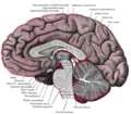 Median sagittal section of brain.
 Pineal gland


[edit] References
- ^ Macchi M, Bruce J (2004). "Human pineal physiology and functional significance of melatonin". Front Neuroendocrinol 25 (3–4): 177–95. doi:10.1016/j.yfrne.2004.08.001. PMID 15589268.
- ^ Arendt J, Skene DJ (2005). "Melatonin as a chronobiotic". Sleep Med Rev 9 (1): 25–39. doi:10.1016/j.smrv.2004.05.002. PMID 15649736. "Exogenous melatonin has acute sleepiness-inducing and temperature-lowering effects during 'biological daytime', and when suitably timed (it is most effective around dusk and dawn) it will shift the phase of the human circadian clock (sleep, endogenous melatonin, core body temperature, cortisol) to earlier (advance phase shift) or later (delay phase shift) times."
- ^ Bowen, R.. "The Pineal Gland and Melatonin". http://www.vivo.colostate.edu/hbooks/pathphys/endocrine/otherendo/pineal.html. Retrieved 14 October 2011.
- ^ a b Zimmerman, Robert A. "Age-Related Incidence of Pineal Calcification Detected by Computed Tomography". Radiological Society of North America. http://radiology.rsna.org/content/142/3/659.full.pdf. Retrieved 21 June 2012.
- ^ Luke, Jennifer. "Fluoride Deposition in the Aged Human Pineal Gland". School of Biological Sciences, University of Surrey, Guildford, UK Department of Obstetrics and Gynaecology, The Royal London Hospital. http://www.icnr.com/articles/fluoride-deposition.html. Retrieved 9 August 2012.
- ^ Kleinschmidt-DeMasters BK, Prayson RA (November 2006). "An algorithmic approach to the brain biopsy—part I". Arch. Pathol. Lab. Med. 130 (11): 1630–8. doi:10.1043/1543-2165(2006)130[1630:AAATTB]2.0.CO;2. PMID 17076524.
- ^ Bocchi G, Valdre G (1993). "Physical, chemical, and mineralogical characterization of carbonate-hydroxyapatite concretions of the human pineal gland". J Inorg Biochem 49 (3): 209–20. doi:10.1016/0162-0134(93)80006-U. PMID 8381851.
- ^ Baconnier S, Lang S, Polomska M, Hilczer B, Berkovic G, Meshulam G (2002). "Calcite microcrystals in the pineal gland of the human brain: first physical and chemical studies". Bioelectromagnetics 23 (7): 488–95. doi:10.1002/bem.10053. PMID 12224052.
- ^ "IngentaConnect High Accumulation of Calcium and Phosphorus in the Pineal Bodies". Ingentaconnect.com. 2006-06-16. http://www.ingentaconnect.com/content/hum/bter/2007/00000119/00000002/art00004. Retrieved 2009-07-06.
- ^ Moore RY, Heller A, Wurtman RJ, Axelrod J (January 1967). "Visual pathway mediating pineal response to environmental light". Science 155 (759): 220–3. doi:10.1126/science.155.3759.220. PMID 6015532.
- ^ a b Schwab, I.R.; O'Connor, G.R. (March 2005). "The lonely eye" (Full text). British Journal of Ophthalmology 89 (3): 256. doi:10.1136/bjo.2004.059105. PMC 1772576. PMID 15751188. //www.ncbi.nlm.nih.gov/pmc/articles/PMC1772576/.
- ^ Pritchard, Thomas C.; Alloway, Kevin Douglas (1999) (Google books preview). Medical Neuroscience. Hayes Barton Press. pp. 76–77. ISBN 1-889325-29-5. http://books.google.com/?id=m7Y80PcFHtsC&printsec=frontcover#PPA76,M1. Retrieved 2009-02-08.
- ^ Kurochkin, Evgeny N.; Gareth J. Dyke, Sergei V. Saveliev, Evgeny M. Pervushov, Evgeny V. Popov (June 2007). "A fossil brain from the Cretaceous of European Russia and avian sensory evolution" (Full text). Biology Letters (The Royal Society) 3 (3): 309–313. doi:10.1098/rsbl.2006.0617. PMC 2390680. PMID 17426009. //www.ncbi.nlm.nih.gov/pmc/articles/PMC2390680/.
- ^ Lerner AB, Case JD, Takahashi Y (1960). "Isolation of melatonin and 5-methoxyindole-3-acetic acid from bovine pineal glands". J Biol Chem 235: 1992–7. PMID 14415935.
- ^ Coates, Paul M. (2005). Encyclopedia of Dietary Supplements. Marc R. Blackman, Gordon M. Cragg, Mark Levine, Joel Moss, Jeffrey D. White. CRC Press. p. 457. ISBN 0-8247-5504-9. http://books.google.com/?id=Sfmc-fRCj10C&pg=PA457&lpg=PA457&dq=Lerner+melatonin+history. Retrieved 2009-03-31.
- ^ Axelrod J (1970). "The pineal gland". Endeavour 29 (108): 144–8. PMID 4195878.
- ^ Lack of pineal growth during childhood. Schmidt F, Penka B, Trauner M, Reinsperger L, Ranner G, Ebner F, Waldhauser F. J Clin Endocrinol Metab. 1995 Apr;80(4):1221–5.
- ^ Development of the pineal gland: measurement with MR. Sumida M, Barkovich AJ, Newton TH. AJNR Am J Neuroradiol. 1996 Feb;17(2):233–6.
- ^ Tapp E, Huxley M. The weight and degree of calcification of the pineal gland. J Pathol 1971;105:31–39
- ^ Tapp E, Huxley M. The histological appearance of the human pineal gland from puberty to old age. J Pathol 1972;108:137–144
- ^ "The Pineal Body". Human Anatomy (Gray's Anatomy). http://www.theodora.com/anatomy/the_pineal_body.html. Retrieved 2011-09-07.
- ^ Strassman
- ^ Natesan A, Geetha L, Zatz M (2002). "Rhythm and soul in the avian pineal". Cell Tissue Res 309 (1): 35–45. doi:10.1007/s00441-002-0571-6. PMID 12111535.
- ^ Uz T, Akhisaroglu M, Ahmed R, Manev H (2003). "The pineal gland is critical for circadian Period1 expression in the striatum and for circadian cocaine sensitization in mice". Neuropsychopharmacology 28 (12): 2117–23. doi:10.1038/sj.npp.1300254. PMID 12865893.
- ^ Uz T, Dimitrijevic N, Akhisaroglu M, Imbesi M, Kurtuncu M, Manev H (2004). "The pineal gland and anxiogenic-like action of fluoxetine in mice". Neuroreport 15 (4): 691–4. doi:10.1097/00001756-200403220-00023. PMID 15094477.
- ^ Manev H, Uz T, Kharlamov A, Joo J (1996). "Increased brain damage after stroke or excitotoxic seizures in melatonin-deficient rats". FASEB J 10 (13): 1546–51. PMID 8940301.
- ^ a b Strassman, Rick J. (2001). DMT: The Spirit Molecule. A Doctor's Revolutionary Research into the Biology of Near-Death and Mystical Experiences. Rochester, Vt: Park Street. ISBN 978-0-89281-927-0. ("Chapter summaries". http://rickstrassman.com/index.php?option=com_content&view=article&id=61&Itemid=60. Retrieved 27 February 2012.)
- ^ Hancock, Graham (2005). Supernatural: Meetings with the Ancient Teachers of Mankind. London: Century. ISBN 978-1-84413-681-0.
- ^ a b Descartes and the Pineal Gland (Stanford Encyclopedia of Philosophy)
- ^ Descartes R. "The Passions of the Soul" excerpted from "Philosophy of the Mind," Chalmers, D. New York: Oxford University Press, Inc.; 2002. ISBN 978-0-19-514581-6
- ^ http://en.wikisource.org/wiki/Ethics_%28Spinoza%29/Part_5
- ^ Hollier, D, Against Architecture: The Writings of Georges Bataille, trans. Betsy Wing, MIT, 1989.
- ^ Bataille, G, Visions of Excess: Selected Writings, 1927–1939 (Theory and History of Literature, Vol 14), trans. Allan Stoekl et al., Manchester University Press, 1985
[edit] External links
| Look up pineal gland in Wiktionary, the free dictionary. |
| Wikimedia Commons has media related to: Pineal gland |
- NeuroNames hier-280
- Histology at BU: Endocrine System: pineal gland (illustration)
- Anatomy Atlases, Microscopic atlas: Pineal gland
- MedPix: Images of Pineal region
- Ancient Brain Mapping and Origins of the Eye of Horus: Egyptian Study of the Pineal Gland
| ||
| ||
| ||


No comments:
Post a Comment